touchINFECTIOUS DISEASES touchINFECTIOUS DISEASES
COVID-19
Read Time: 6 mins

Confirmed SARS-CoV-2 Reinfection After 1 Year in a Patient with X-linked Agammaglobulinaemia

Copy Link
Published Online: Aug 30th 2022 touchREVIEWS in Infectious Diseases. 2022;1(1):35-8 DOI: https://doi.org/10.17925/ID.2022.1.1.35
Authors: Gereon J Rieke, Malte B Monin, Sven Breitschwerdt, Christoph Boesecke, Stefan Schlabe
Quick Links:
Abstract
Article
Article Information
Abstract:
Overview

Though a comprehensive analysis of the immunity following severe acute respiratory syndrome coronavirus 2 (SARS-CoV-2) infection has been performed, little is known about the duration of this protection and the risk of reinfection. This lack of knowledge is of particular interest for patients with impaired immune function. In this report, we describe the course of infection of a 30-year-old male patient with X-linked agammaglobulinaemia, who was reinfected with SARS-CoV-2 after a primary infection 12 months earlier. The initial course of infection took place in April 2020 with the typical symptoms of an upper respiratory tract infection accompanied by compatible changes in laboratory values and computed tomography. With no anti-viral treatment options at that time of the pandemic, only symptomatic therapy could be offered. Twelve months later (April 2021), the patient presented with a short course of fever and headache. Laboratory testing showed elevated C-reactive protein levels, while leukocytes, lymphocytes and lactate dehydrogenase levels were within range. The patient was admitted, and antibiotic treatment was started partially because procalcitonin levels were slightly elevated as well. The SARS-CoV-2 polymerase chain reaction was positive, and therapy with the monoclonal SARS-CoV-2 antibodies casirivimab/imdevimab (1,200 mg/1,200 mg, respectively) were initiated. The course of infection was mild, but low-flow oxygen had to be administered. It was not possible to distinguish between the contribution of the administered antibodies and the role of cytotoxic T-cells in the course of infection. Variant screenings confirmed the Wuhan strain of the virus for the first episode and the alpha variant for the second episode, thus confirming reinfection and ruling out long-term shedding. Neutralizing antibodies seem to play a crucial role in viral clearance and infection prevention, assuming patients with agammaglobulinaemia are at higher risk for a severe course of coronavirus disease 2019. Still, the specific role of neutralizing antibodies and cytotoxic T-cells is not fully understood. Reinfection among this patient population has only been described occasionally. Our case described a reinfection, which was confirmed by variant-testing. In addition, it gave insight into the rapid progression of testing and into specific anti-viral therapy over 1 year of the pandemic.

Keywords

Case report, coronavirus diease 2019, reinfection, severe acute respiratory syndrome coronavirus 2,
X-linked agammaglobulinaemia

Article:

With the on-going coronavirus disease 2019 (COVID-19) pandemic challenging almost every aspect of life, immunity to COVID-19 following vaccination or infection has become the centre of attention. Several vaccines provide protection, but the duration of protective immunity is not known. Even less understood are the duration of protection after natural infection and the role of different immune cells.1 Specific interest has been given to patients with impaired immune function due to primary immunodeficiencies, HIV, immunosuppressive medication or immunosenescence. Here we present the disease course of a patient with X-linked agammaglobulinaemia (XLA) following infection and confirmed reinfection after 1 year with different SARS-CoV-2 virus strains before the onset of the omicron wave.

Case report

This 30-year-old male patient with XLA had a history of multiple infectious diseases. He was receiving weekly subcutaneous immunoglobulins (SCIG). In April 2020, he presented with a 10-day history of fever and abdominal pain, dyspnoea and shortness of breath. His respiratory rate was 20 breaths/minute, and he had an oxygen saturation of 90% on room air. The SARS-CoV-2 infection was confirmed by polymerase chain reaction (PCR) in an oropharyngeal swab (see Supplemental Table 1 for the methods and kits). He received oxygen supplementation via a nasal cannula.

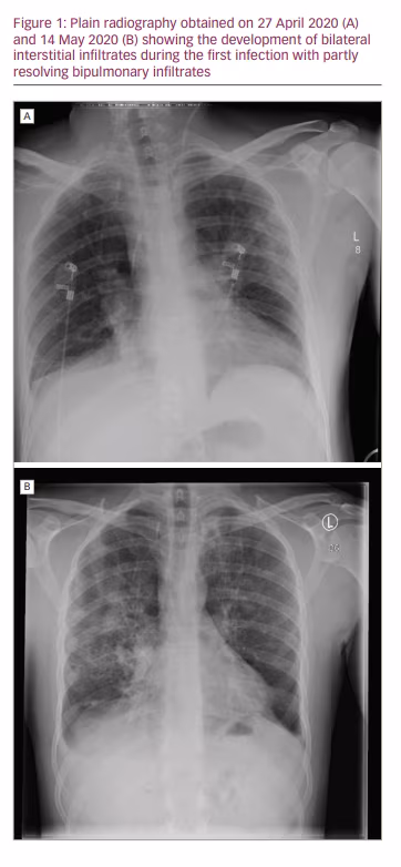
Initially, C-reactive protein (CRP) was 64.7 mg/L (normal values: <5.0 mg/L) and peaked at 170 mg/L (day 10). Leukocytes and lymphocytes were normal, and platelet count was 91 × 109/L (normal value: 150–370 × 109/L), with a minimum of 81 × 109/L (day 2). Immunoglobulins were tested once, and a value of 3.99 g/L (normal values: 7.0–16.0 g/L) was found during peak inflammation. Chest X-ray initially showed no signs of pneumonia (Figure 1A) but did later in the disease course (Figure 1B).

The administration of convalescent plasma was discussed but dismissed due to only mild symptoms at that time and fear of severe inflammation via an antibody-enhanced inflammatory response, which would worsen the clinical status. Since no specific anti-viral medication was available, the patient received intravenous fluids, prophylactic
low-molecular-weight heparin and anti-pyretics.

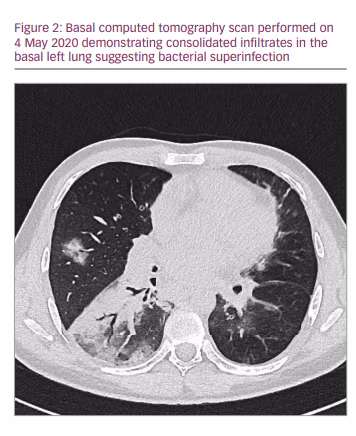
A computed tomography scan showed ground-glass opacification and infiltrates assigned to bacterial superinfection (Figure 2). After defervescence and ceasing supplemental oxygen on day 19, he could be discharged on day 23. He was symptom free and tested negative for SARS-CoV-2 via PCR multiple times. Enzyme-linked immunosorbent assays (ELISAs) for anti-SARS-CoV-2 antibodies proved inconclusive at the time of discharge. One ELISA showed signs of immunoglobulin (Ig) G, and two other ELISAs detected neither IgG nor IgA.

Precisely 1 year later, in April 2021, the patient presented again with fever and concomitant headache developed the previous day. He had received a dose of SCIG the day the fever had started. PCR was positive for SARS-CoV-2. Further testing revealed deletion 69/70 and N501Y, classifying the virus as the variant of concern (VOC-202012/01) or B.1.1.7, later assigned as the alpha variant (see Supplemental Table 1 for further details).

Leukocytes, lymphocytes and lactate dehydrogenase were normal, while the platelet count was slightly decreased (121 × 109/L), and the CRP was elevated (49.4 mg/L rising to 154.4 mg/L). Procalcitonin was initially slightly elevated at 0.57 µg/L (normal values: <0.50 µg/L). The patient was admitted to the hospital and received empiric antibiotic treatment, intravenous fluids, anti-pyretics and low-molecular-weight heparin. After confirming the SARS-CoV-2 infection, monoclonal SARS-CoV-2 antibodies casirivimab (1,200 mg) and imdevimab (1,200 mg) (Regeneron Pharmaceuticals, Rensselaer, NY, USA) were administered. We decided to use the monoclonal antibodies first instead of remdesivirs because we expected that they would replace the missing physiological antibody response.

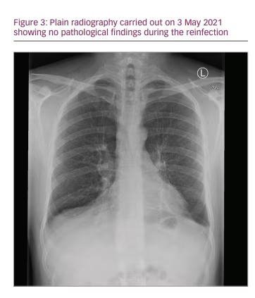
Signs of hyperinflammation, apart from the markedly elevated CRP, were mostly clinical. While the patient was in a better condition than in the first episode, he was still very sick with a high fever. The fever resolved, and no supplemental oxygen or further anti-viral medication was needed. Since low levels of bacteria, Campylobacter jejuni, were discovered in the blood cultures, the patient was kept in the hospital for further observation and intravenous antibiotic treatment for 7 days. He was discharged free of symptoms, with two negative PCR tests and no signs of infiltration on a chest X-ray at 10 days (Figure 3). Testing for antibodies again proved inconclusive. No antibodies against SARS-CoV-2 nucleocapsid protein were detected, but tests for anti-SARS-CoV-2 spike IgM and IgG were positive; confounding treatment with monoclonal anti-spike IgG1 and SCIG should be taken into account.

To rule out possibilities other than reinfection, such as prolonged viral shedding, we performed a variant screening on the stored samples from the initial infection. In this sample, no variants of concern were discovered, suggesting that it was the Wuhan strain.

Discussion

The role of neutralizing antibodies in the viral clearance and prevention of SARS-CoV-2 infection and reinfection has become a main focus since the beginning of the pandemic. Moreover, antibody concentration is of particular interest when evaluating the efficacy of vaccinations and booster campaigns. Therefore, patients with immunodeficiency have been initially regarded as at risk for a severe course of COVID-19.2 The natural course, however, and the specific benefit of the available therapies, especially convalescent plasma, in these patients are still unknown.

So far, 18 case reports and small series of a total of 32 patients with XLA contracting COVID-19 have been published.3–20 Most patients had a mild course of COVID-19 and were able to clear the virus. Initially, this suggested that antibodies may not be decisive for clearing the disease in patients with functional cytotoxic T-cells. Moreover, it was speculated that the lack of B-cell-derived cytokines, such as interleukin-6 and tumour necrosis factor-α, may prevent the ‘cytokine storm’ seen in severe COVID-19.7,8,16 Recently, reports on prolonged and severe disease courses, including one deceased patient, have been published.4,5,12,18 They show that there are indeed patients with severe inflammation. Only one patient, however, needed mechanical ventilation.12 Thirteen of these 32 patients received convalescent plasma at varying time points, often after several weeks of illness. Most patients recovered quickly thereafter; no unfavourable outcomes associated with convalescent plasma were reported.21

Even without a clear benefit being shown in randomized studies, some authors argued that, in a specific group of patients with COVID-19, such as those with primary immunodeficiencies, viral clearance may depend on neutralizing antibodies provided by convalescent plasma.22 While we decided against its administration during our patient’s first infection due to mild disease, convalescent plasma as a therapeutic option had been largely replaced by monoclonal antibodies at the time of his second infection. Anti-spike IgG1 antibodies such as imdevimab and casirivimab were broadly introduced to prevent hospitalization and severe disease in patients in the early phase of the SARS-CoV-2 infection.23 In our case, the second infection was milder and shorter than the first. We can, however, not decide about the contribution of monoclonal antibody administration to the observed mild disease course or, indeed, the role of cytotoxic T-cells or the virus variants.

Most importantly, the specific role of neutralizing antibodies and cytotoxic T-cells in clearing the virus and preventing reinfection generally and in patients with XLA particularly is unclear. Reinfection with SARS-CoV-2, first reported in the summer of 2020,24 at this time was rare given the numbers of infected patients. Clinical characteristics, duration and course of reinfections, as well as risk factors, have not been defined yet. To the best of our knowledge, the patient presented here was the first confirmed patient with XLA and SARS-CoV-2 reinfection. There is only one case report on a suspected reinfection less than 3 weeks after initial recovery in a patient with XLA. This published case does show a possible but not undoubtedly proven reinfection, given the short time between the first and presumed second episode.5 The short time, however, does not exclude reinfection. Long-term shedding, as described in several patients with XLA,12,18 or recrudescence and a second peak of illness as also described,18 could not be ruled out as no sequencing was performed. In our case, reinfection was undoubtedly confirmed by variant screening, which revealed infection with a different variant after 1 year, highlighting that these patients indeed face the risk of reinfection. Apart from specific aspects in patients with primary immunodeficiency, this case illustrates the impressive progress in diagnostics and therapy over 1 year of the COVID-19 pandemic.

Article Information:
Disclosure

Gereon J Rieke had a scholarship from the University of Bonn (BONFOR). He received payment for an interview for Gilead about remdesivir (€450). Christoph Boesecke received academic funding from NEAT ID, Hector Stiftung and the German Centre for Infectious Diseases Research (DZIF); he received consulting fees and payment or honoraria for lectures, presentations, speakers’ bureaus, manuscript writing or educational events from AbbVie, Gilead, Janssen, MSD and ViiV. He states a leadership or fiduciary role in other board, society, committee or advocacy groups, paid or unpaid, in DAIG and EACS (board member). Stefan Schlabe received a stipend from the DZIF. He received congress fees and travel expenses not related to the topic of the case report from Gilead, AbbVie and Johnson & Johnson. He holds shares in BioNTech (fonds based, MIG9). Malte B Monin and Sven Breitschwerdt have no financial or non-financial relationships or activities to declare in relation to this article.

Compliance With Ethics

All procedures were followed in accordance with the responsible committee on human experimentation and with the Helsinki Declaration of 1975 and subsequent revisions, and informed consent was received from the patient involved in this case study.

Review Process

Double-blind peer review.

Authorship

The named authors meet the International Committee of Medical Journal Editors (ICMJE) criteria for authorship of this manuscript, take responsibility for the integrity of the work as a whole, and have given final approval for the version to be published.

Correspondence

Gereon J Rieke, Department of Medicine I, University Hospital of Bonn, 53127 Bonn, Germany. E: Gereon.Rieke@ukbonn.de

Support

No funding was received in the publication of this article.

Access

This article is freely accessible at touchINFECTIOUSDISEASES.com. © Touch Medical Media 2022

Acknowledgements

We thank the Institute of Virology in Bonn for variant screening and the Department of Radiology for providing the images.

Data Availability

Data sharing is not applicable to this article as no datasets were generated or analysed during the writing of this study.

Received

2022-06-15

References

  1. Khoshkam Z, Aftabi Y, Stenvinkel P, et al. Recovery scenario and immunity in COVID-19 disease: A new strategy to predict the potential of reinfection. J Adv Res. 2021;31:49–60.
  2. Mohammed AH, Blebil A, Dujaili J, Rasool-Hassan BA. The risk and Impact of COVID-19 pandemic on immunosuppressed patients: Cancer, HIV, and solid organ transplant recipients. AIDS Rev. 2020;22:151–7.
  3. Hovey JG, Tolbert D, Howell D. Bruton’s agammaglobulinemia and COVID-19. Cureus. 2020;12:e11701.
  4. Iaboni A, Wong N, Betschel SD. A patient with X-linked agammaglobulinemia and COVID-19 infection treated with remdesivir and convalescent plasma. J Clin Immunol. 2021;41:923–5.
  5. Loh SY, Bassett J, Hoodless EJ, Walshaw M. Possible COVID-19 reinfection in a patient with X-linked agammaglobulinaemia. BMJ Case Rep. 2021;14:e240765.
  6. Milosevic I, Jovanovic J, Stevanovic O. Atypical course of COVID-19 in patient with Bruton agammaglobulinemia. J Infect Dev Ctries. 2020;14:1248–51.
  7. Mira E, Yarce OA, Ortega C, et al. Rapid recovery of a SARS-CoV-2-infected X-linked agammaglobulinemia patient after infusion of COVID-19 convalescent plasma. J Allergy Clin Immunol Pract. 2020;8:2793–5.
  8. Meyts I, Bucciol G, Quinti I, et al. Coronavirus disease 2019 in patients with inborn errors of immunity: An international study. J Allergy Clin Immunol. 2021;147:520–31.
  9. Quinti I, Lougaris V, Milito C, et al. A possible role for B cells in COVID-19? Lesson from patients with agammaglobulinemia. J Allergy Clin Immunol. 2020;146:211–3.e4.
  10. Soresina A, Moratto D, Chiarini M, et al. Two X-linked agammaglobulinemia patients develop pneumonia as COVID-19 manifestation but recover. Pediatr Allergy Immunol. 2020;31:565–9.
  11. Ho HE, Mathew S, Peluso MJ, Cunningham-Rundles C. Clinical outcomes and features of COVID-19 in patients with primary immunodeficiencies in New York City. J Allergy Clin Immunol Pract. 2021;9:490–3.e2.
  12. Guetl K, Moazedi-Fuerst F, Rosskopf K, et al. SARS-CoV-2 positive virus culture 7 weeks after onset of COVID-19 in an immunocompromised patient suffering from X chromosome-linked agammaglobulinemia. J Infect. 2021;82:414–51.
  13. Shields AM, Burns SO, Savic S, Richter AG. COVID-19 in patients with primary and secondary immunodeficiency: The United Kingdom experience. J Allergy Clin Immunol. 2021;147:870–5.e1.
  14. Almontasheri A, Al-Husayni F, Alsuraihi AK, et al. The clinical course of COVID-19 pneumonia in a 19-year-old man on intravenous immunoglobulin replacement therapy for X-linked agammaglobulinemia. Am J Case Rep. 2021;22:e929447.
  15. Jin H, Reed JC, Liu STH, et al. Three patients with X-linked agammaglobulinemia hospitalized for COVID-19 improved with convalescent plasma. J Allergy Clin Immunol Pract. 2020;8:3594–6.e3.
  16. Marcus N, Frizinsky S, Hagin D, et al. Minor clinical impact of COVID-19 pandemic on patients with primary immunodeficiency in Israel. Front Immunol. 2021;11:614086.
  17. Delavari S, Abolhassani H, Abolnezhadian F, et al. Impact of SARS-CoV-2 pandemic on patients with primary immunodeficiency. J Clin Immunol. 2021;41:345–55.
  18. Buckland MS, Galloway JB, Fhogartaigh CN, et al. Treatment of COVID-19 with remdesivir in the absence of humoral immunity: A case report. Nat Commun. 2020;11:6385.
  19. Delgado-Fernandez M, Garcia-Gemar GM, Fuentes-Lopez A, et al. Treatment of COVID-19 with convalescent plasma in patients with humoral immunodeficiency – Three consecutive cases and review of the literature. Enferm Infecc Microbiol Clin (Engl Ed). doi: 10.1016/j.eimc.2021.01.013.
  20. Pereira NMD, Heath PT, Doerholt K, et al. SARS-CoV-2 infection in an adolescent with X-linked agammaglobulinemia. Pediatr Infect Dis J.2021;40:e472–e4.
  21. Lang-Meli J, Fuchs J, Mathe P, et al. Case series: Convalescent plasma therapy for patients with COVID-19 and primary antibody deficiency. J Clin Immunol. 2022;42:253–65.
  22. Rnjak D, Šola AM, Halassy B, et al. COVID-19 convalescent plasma as long-term therapy in immunodeficient patients? Transfus Clin Biol. 2021;28:264–70.
  23. Weinreich DM, Sivapalasingam S, Norton T, et al. REGN-COV2, a neutralizing antibody cocktail, in outpatients with Covid-19. N Engl J Med. 2021;384:238–51.
  24. Bongiovanni M, Basile F. Re-infection by COVID-19: A real threat for the future management of pandemia? Infect Dis (Lond). 2020;522:581–2.

Further Resources

Share this Article
Related Content In COVID-19
  • Copied to clipboard!
    accredited arrow-down-editablearrow-downarrow_leftarrow-right-bluearrow-right-dark-bluearrow-right-greenarrow-right-greyarrow-right-orangearrow-right-whitearrow-right-bluearrow-up-orangeavatarcalendarchevron-down consultant-pathologist-nurseconsultant-pathologistcrosscrossdownloademailexclaimationfeedbackfiltergraph-arrowinterviewslinkmdt_iconmenumore_dots nurse-consultantpadlock patient-advocate-pathologistpatient-consultantpatientperson pharmacist-nurseplay_buttonplay-colour-tmcplay-colourAsset 1podcastprinter scenerysearch share single-doctor social_facebooksocial_googleplussocial_instagramsocial_linkedin_altsocial_linkedin_altsocial_pinterestlogo-twitter-glyph-32social_youtubeshape-star (1)tick-bluetick-orangetick-red tick-whiteticktimetranscriptup-arrowwebinar Sponsored Department Location NEW TMM Corporate Services Icons-07NEW TMM Corporate Services Icons-08NEW TMM Corporate Services Icons-09NEW TMM Corporate Services Icons-10NEW TMM Corporate Services Icons-11NEW TMM Corporate Services Icons-12Salary £ TMM-Corp-Site-Icons-01TMM-Corp-Site-Icons-02TMM-Corp-Site-Icons-03TMM-Corp-Site-Icons-04TMM-Corp-Site-Icons-05TMM-Corp-Site-Icons-06TMM-Corp-Site-Icons-07TMM-Corp-Site-Icons-08TMM-Corp-Site-Icons-09TMM-Corp-Site-Icons-10TMM-Corp-Site-Icons-11TMM-Corp-Site-Icons-12TMM-Corp-Site-Icons-13TMM-Corp-Site-Icons-14TMM-Corp-Site-Icons-15TMM-Corp-Site-Icons-16TMM-Corp-Site-Icons-17TMM-Corp-Site-Icons-18TMM-Corp-Site-Icons-19TMM-Corp-Site-Icons-20TMM-Corp-Site-Icons-21TMM-Corp-Site-Icons-22TMM-Corp-Site-Icons-23TMM-Corp-Site-Icons-24TMM-Corp-Site-Icons-25TMM-Corp-Site-Icons-26TMM-Corp-Site-Icons-27TMM-Corp-Site-Icons-28TMM-Corp-Site-Icons-29TMM-Corp-Site-Icons-30TMM-Corp-Site-Icons-31TMM-Corp-Site-Icons-32TMM-Corp-Site-Icons-33TMM-Corp-Site-Icons-34TMM-Corp-Site-Icons-35TMM-Corp-Site-Icons-36TMM-Corp-Site-Icons-37TMM-Corp-Site-Icons-38TMM-Corp-Site-Icons-39TMM-Corp-Site-Icons-40TMM-Corp-Site-Icons-41TMM-Corp-Site-Icons-42TMM-Corp-Site-Icons-43TMM-Corp-Site-Icons-44TMM-Corp-Site-Icons-45TMM-Corp-Site-Icons-46TMM-Corp-Site-Icons-47TMM-Corp-Site-Icons-48TMM-Corp-Site-Icons-49TMM-Corp-Site-Icons-50TMM-Corp-Site-Icons-51TMM-Corp-Site-Icons-52TMM-Corp-Site-Icons-53TMM-Corp-Site-Icons-54TMM-Corp-Site-Icons-55TMM-Corp-Site-Icons-56TMM-Corp-Site-Icons-57TMM-Corp-Site-Icons-58TMM-Corp-Site-Icons-59TMM-Corp-Site-Icons-60TMM-Corp-Site-Icons-61TMM-Corp-Site-Icons-62TMM-Corp-Site-Icons-63TMM-Corp-Site-Icons-64TMM-Corp-Site-Icons-65TMM-Corp-Site-Icons-66TMM-Corp-Site-Icons-67TMM-Corp-Site-Icons-68TMM-Corp-Site-Icons-69TMM-Corp-Site-Icons-70TMM-Corp-Site-Icons-71TMM-Corp-Site-Icons-72